Foot & Ankle
Find relief from your foot and ankle condition with the support of our expert-led multidisciplinary team. We’re committed to providing the highest level of care and supporting you every step of the way.
Quick links

QUICK LINKS
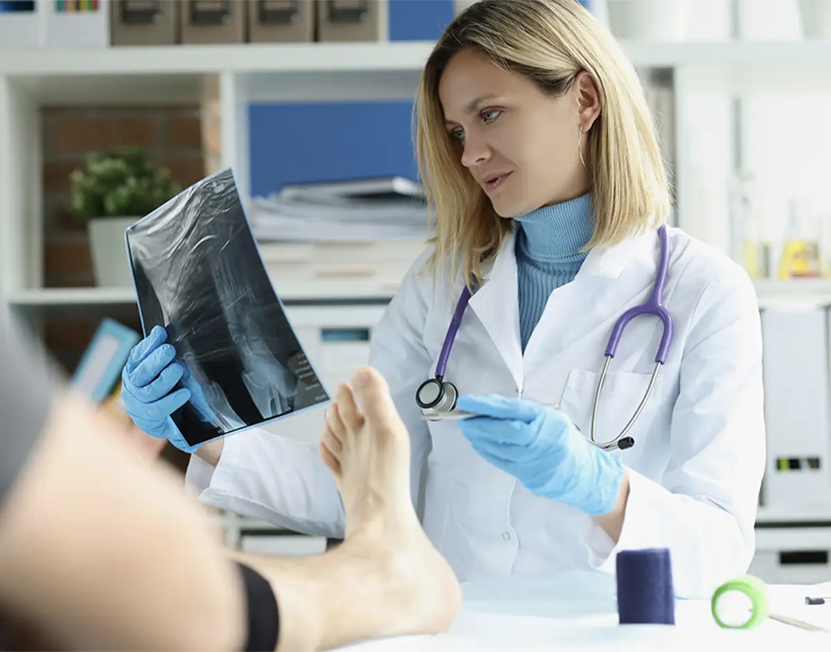
Experts in foot and ankle care
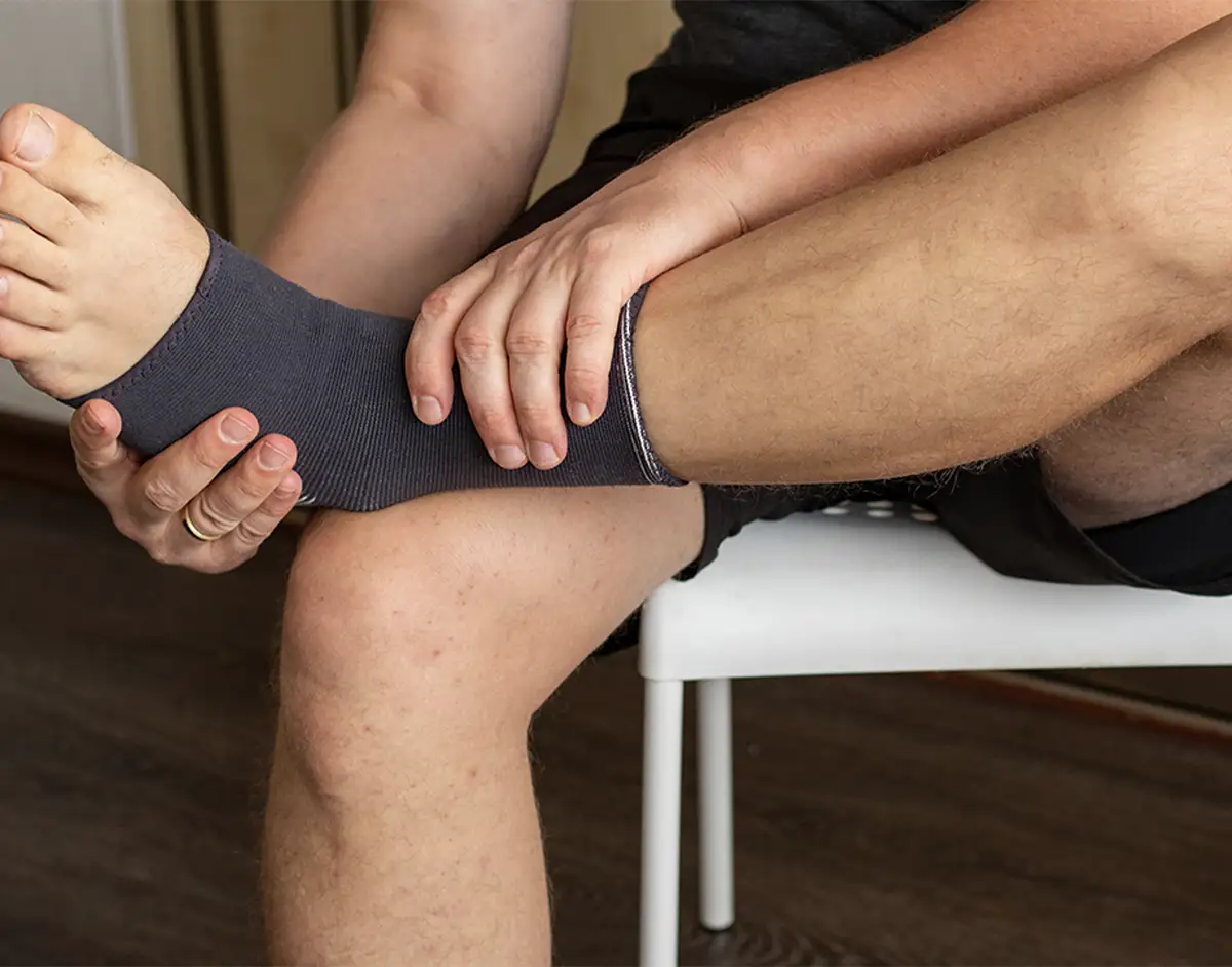
Our expert foot and ankle specialists treat a wide range of lower limb conditions, helping you walk and move freely without pain and troublesome symptoms.
Professor Nima Heidari specialises in managing foot and ankle conditions, including trauma and sports injuries. He is also highly skilled in treating complex fractures and has a special interest in joint preservation for arthritis, enabling you to get back to doing what you love.
Mr Thomas Hester brings an extensive background in research and innovation in foot and ankle conditions, ensuring you receive expert-level care. For younger patients, our paediatric consultants, Mr Thomas Crompton, Ms Claudia Maizen and Mr Kyle James, support children and their families seeking treatment for troublesome foot and ankle conditions.
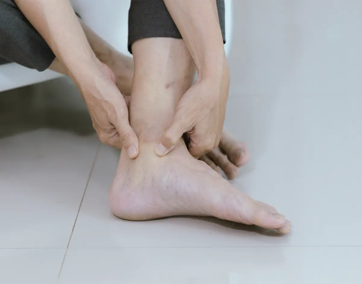
Foot and ankle anatomy
The feet and ankles are highly flexible, complex structures containing multiple bones, tendons and ligaments. Our foot and ankle team treat all conditions ranging from bunions, bone deformity and arthritis to painful ligament and Achilles tendon injuries.
The foot is divided into three parts:
The forefoot consists of five toes (phalanges) and five longer bones (metatarsals).
The midfoot forms the arch and consists of the three cuneiform, cuboid and navicular bones.
The hindfoot forms the heel and ankle. The calcaneus is the heel bone.
The ankle is formed of the talus bone, which supports the tibia (shin bone) and fibula in the leg. The tibiotalar joint (ankle joint) allows the foot to move up and down. The lateral malleolus is the bony protrusion on the outer ankle, formed by the distal end of the fibula. The medial malleolus is the inner ankle bone, formed by the distal end of the tibia.
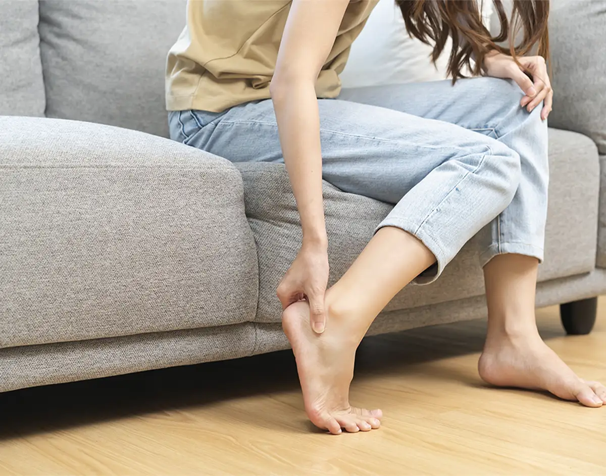
Cartilage cushions the bones and allows them to glide smoothly over one another. Tendons connect muscles to bone to provide support. The Achilles tendon, which wraps around the heel bone, is the largest and strongest tendon in the body. Bursae, small sacs that contain synovial fluid, help to decrease friction between tendons and bones or skin.
Ligaments connect bones to other bones. The plantar fascia is the longest foot ligament, acting as a shock absorber and supporting the foot arch. Other ligaments include the talo-fibular and calcaneo-fibular ligaments.
There are 20 muscles in the foot, categorised as intrinsic (responsible for toe movement) and extrinsic (located in the lower leg and responsible for foot movement).
Foot and ankle services
We also offer specialist care through clinics that focus on diabetic foot care, sports management and paediatrics.
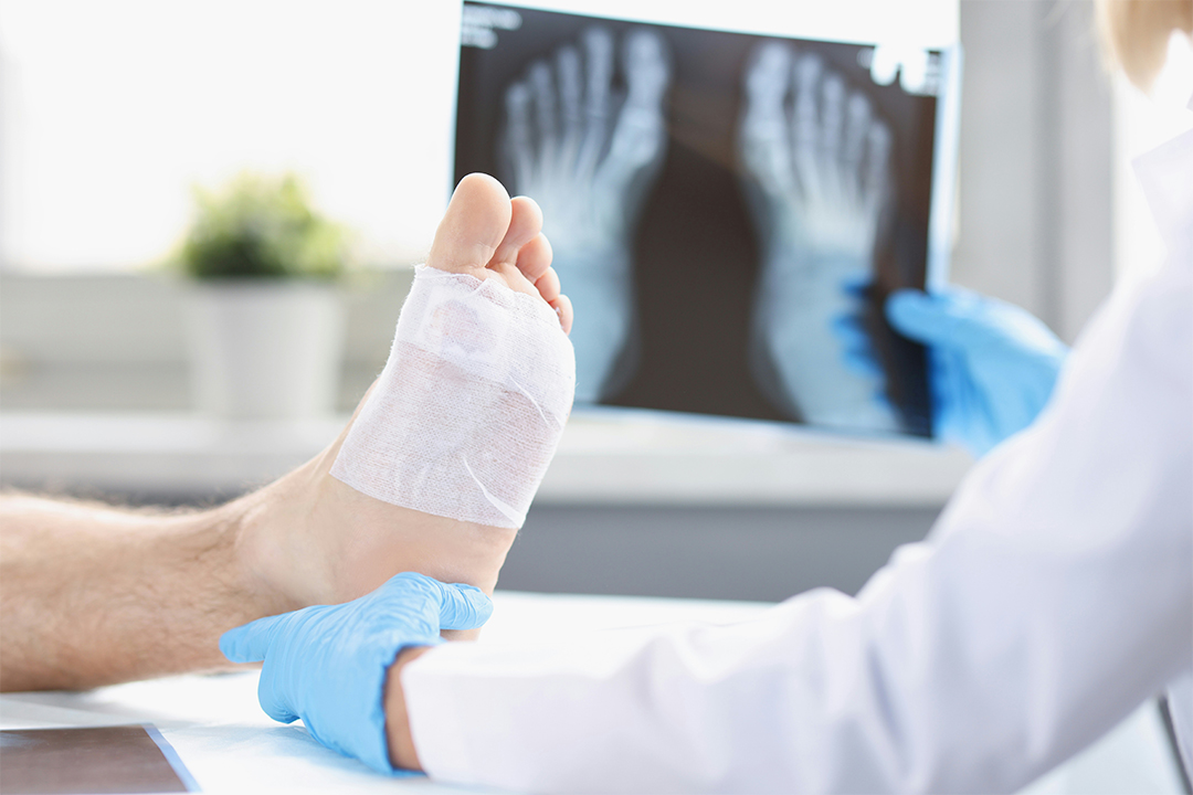
Diabetic Foot Service Clinic
Our Diabetic Foot Service Clinic, led by Professor Nima Heidari, provides patients with expert support in managing their foot health and diabetes.
This clinic offers treatments such as:
● Ulcer debridement
● Bone exostectomy
● Soft tissue balancing
● Minor amputations
● Reconstruction
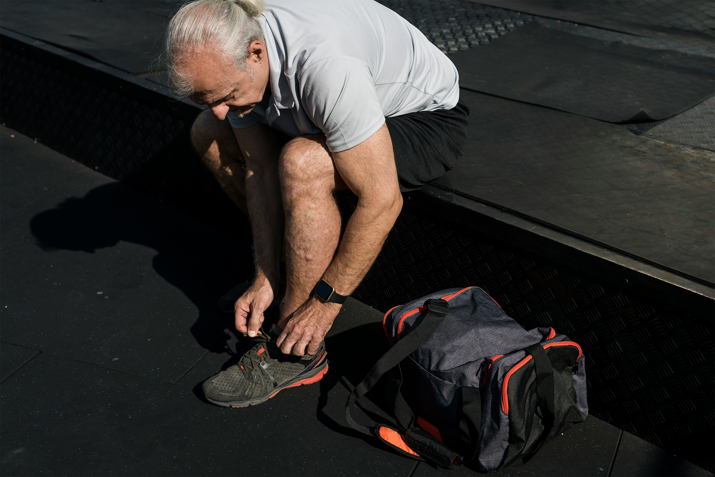
Sports Management Clinic
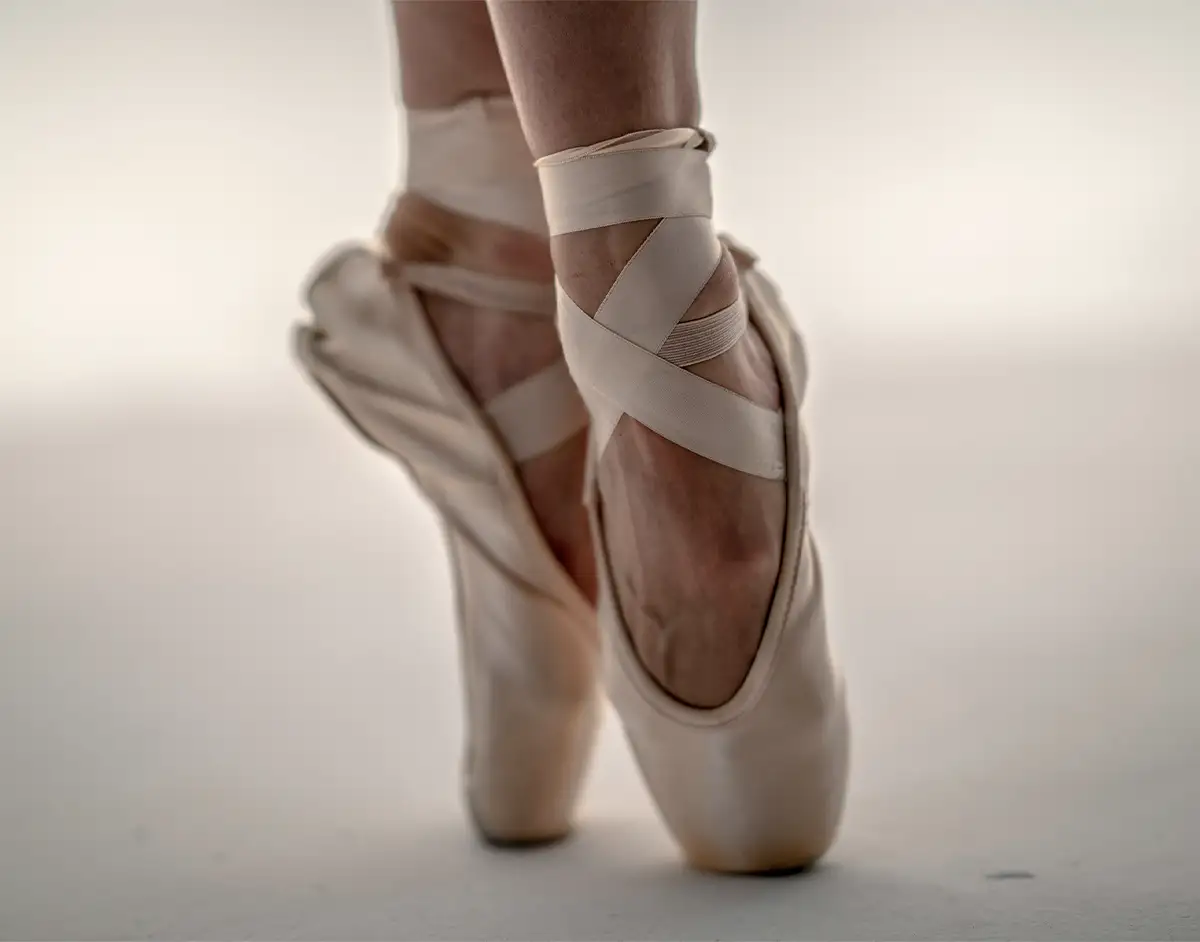
Our Sports Management Clinic is designed for athletes seeking to manage their foot and ankle condition. With experience working with a number of top athletes in gymnastics, ballet, football and running, we’re well-equipped to address a wide range of conditions and injuries that sports can lead to.
We’ll develop a unique treatment plan that helps you to not only recover but also get back to competing. Our team will work with you on preventative strategies to minimise your risk of future injuries too.
Paediatric care
We provide foot and ankle care for all ages, with our team of paediatric consultants ensuring your child receives the care they need. We work with specialist paediatric facilities that help create a comfortable environment, making it easier for your child to attend appointments with us.
Why OS Clinic?
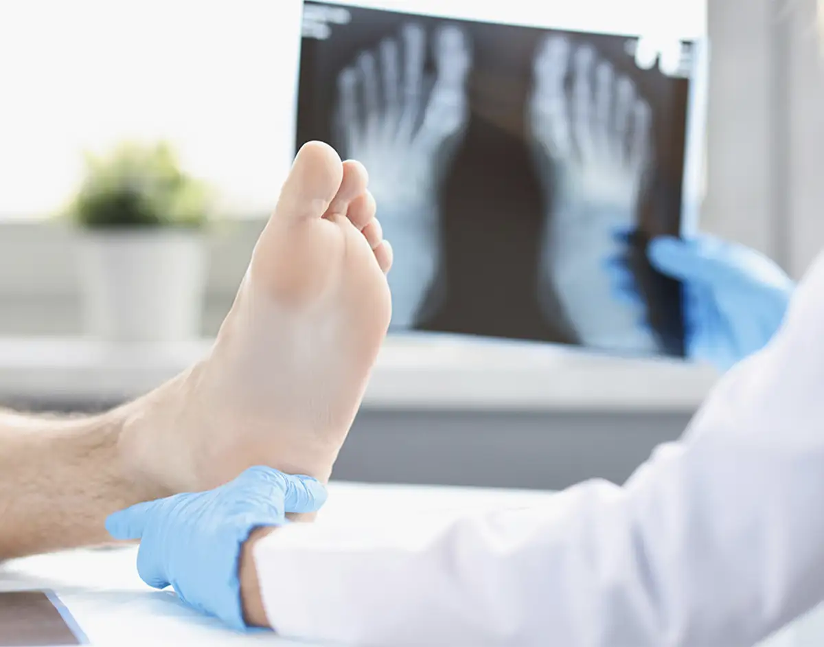
Our team has extensive knowledge of the complex anatomy of the foot and ankle and is skilled in performing minimally invasive surgeries to treat both routine and complex lower limb conditions.
Known internationally, our consultants are leaders in their respective fields, using the latest medical advancements to ensure you receive the very best care. Our multidisciplinary team also focuses on prevention, so you can be confident that not only is your condition treated, but your risk of it returning is minimised.
Our telephone support is available seven days a week so you can always receive the care you need. We also provide same-day diagnostics wherever possible so you can be confident that your healthcare journey is on the right track.
We’re here to help both adults and children find relief from challenging foot and ankle conditions through detailed consultations and tailored treatment plans.