Knee
Our expert team of orthopaedic specialists offers a comprehensive range of knee treatments and is dedicated to providing first-class care to help you get back to living your best life.
Quick links

QUICK LINKS
Experts in the diagnosis and treatment of knee conditions
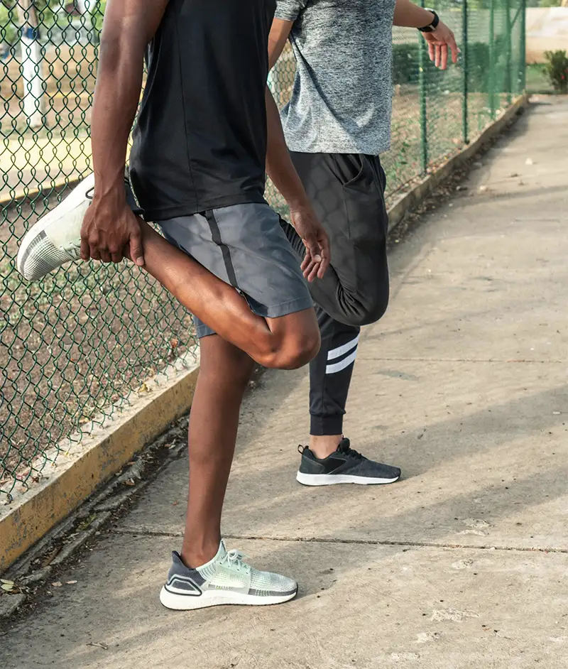
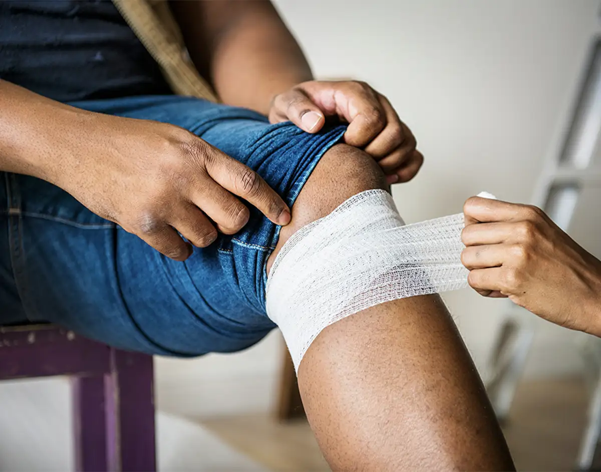
The knee is the largest and most complex joint in your body and can be affected by a wide range of injuries and conditions, each requiring careful attention and care. Our knee consultants are specialists in managing all types of knee issues from traumatic injuries such as ACL rupture to degenerative conditions like arthritis.
Using advanced imaging and diagnostic techniques, our knee specialists accurately diagnose conditions and design highly individualised treatment plans to ensure the best possible outcomes and patient satisfaction. In many cases, our diagnostic tests are often available on the same day as your initial appointment, accelerating your path to treatment and recovery.
Made up of surgeons, musculoskeletal radiologists, physiotherapists, sports medicine specialists and other healthcare professionals, our multidisciplinary knee team works together to provide comprehensive care for knee conditions of all complexities and severity.
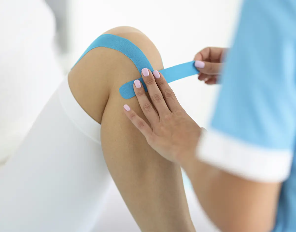
Offering the most advanced treatments, our skilled team provide a wide range of both non-surgical and surgical treatments including physiotherapy, bracing, injections, minimally invasive arthroscopy, total knee replacement, partial knee replacement and complex reconstructions.
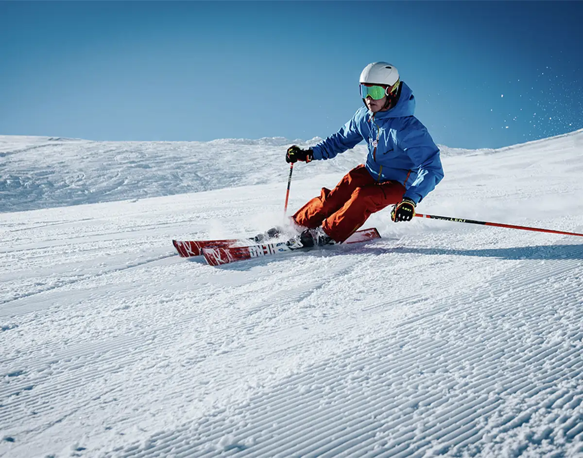
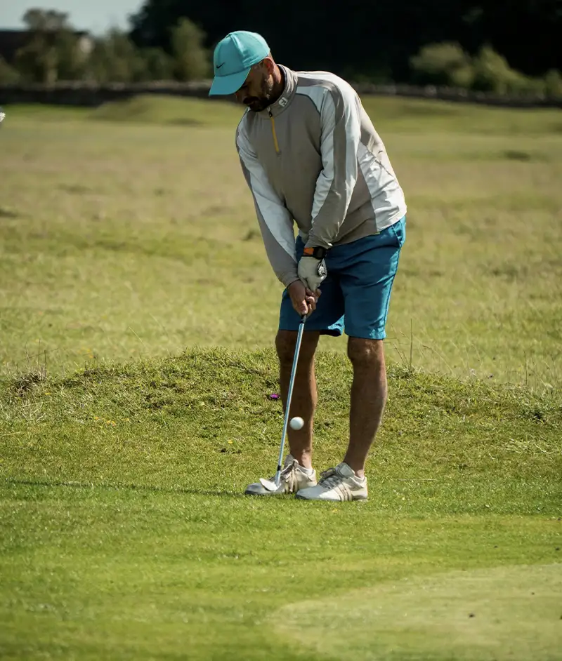
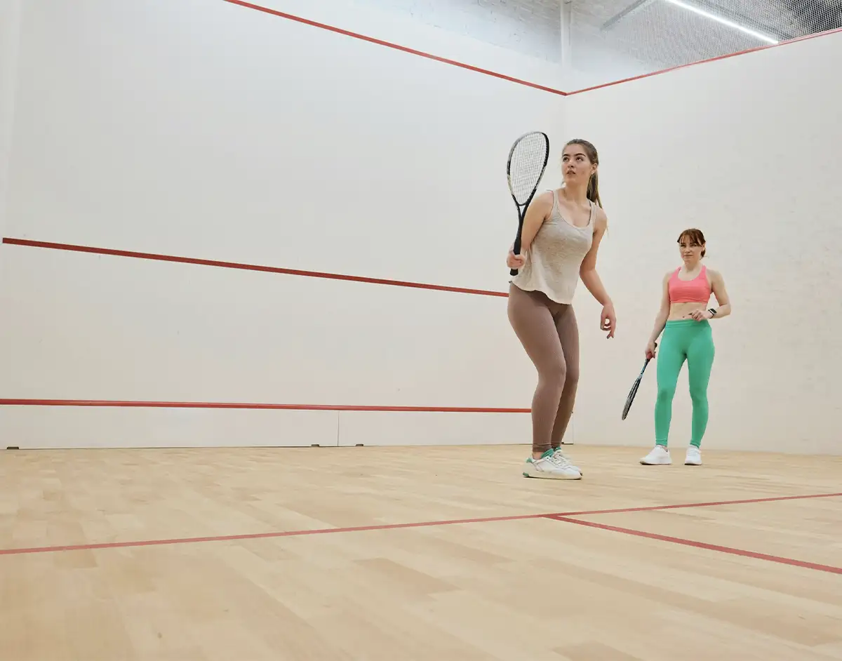
Working closely with our sports and exercise medicine physicians and other orthopaedic consultants with a specific interest in sporting injuries, our knee specialists are also equipped to provide the highest standard of care and condition management to professional and elite athletes with sports injuries.
In our innovative Joint Preservation Clinic our experts offer a range of treatment options to help preserve the knee joint to prevent or delay the need for joint replacement surgery for as long as possible.
Whatever your reason for visiting us, from prehabilitation to treatment to post-surgical rehab, our team are here to provide you with world-class care from the moment you walk through our doors.