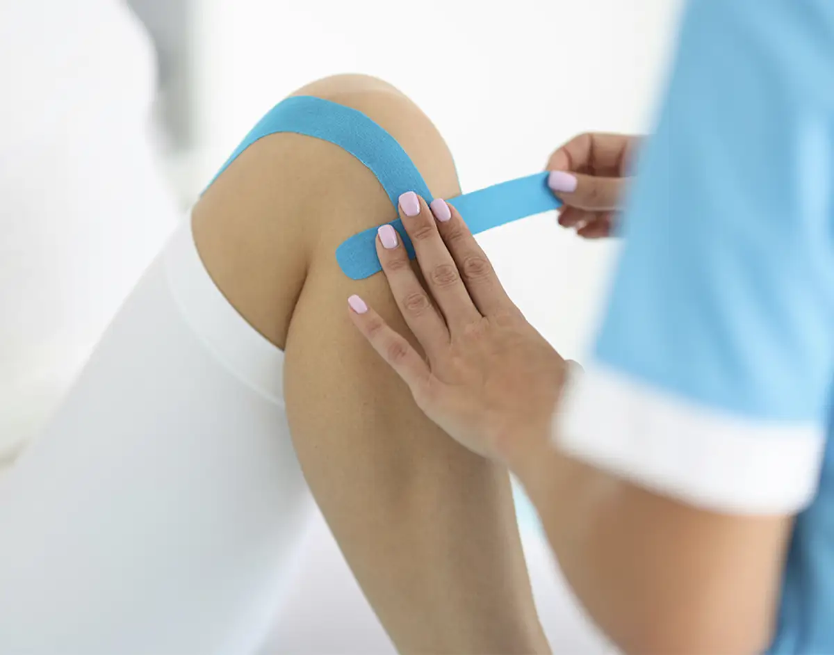
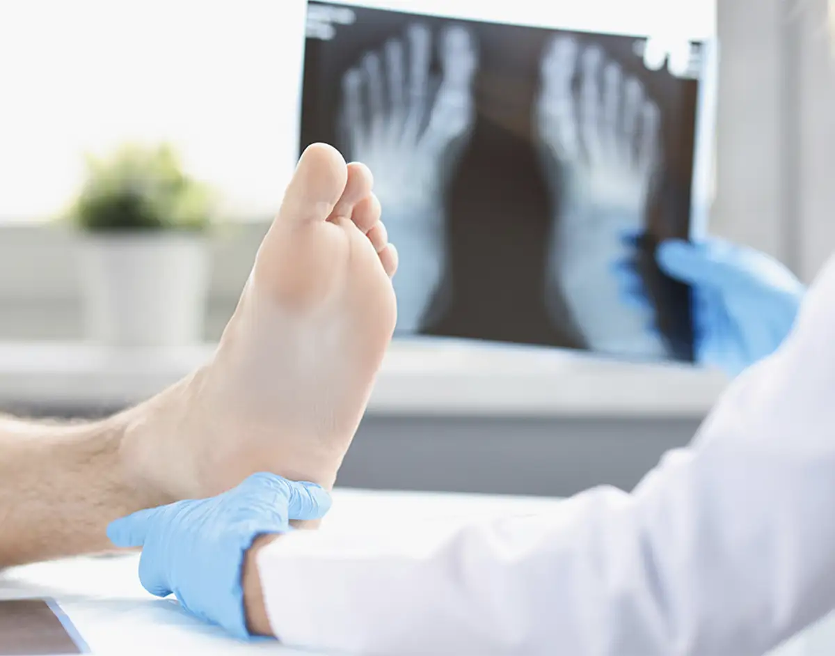
Knee cartilage surgery
Although minor damage to the cartilage may heal by itself, more severe injuries usually need surgical treatment. This is normally carried out using keyhole surgery as a day case under a light general anaesthetic. In more complex cases, other procedures may be advised as well. These include knee realignment surgery (osteotomy).

QUICK LINKS
Knee cartilage surgery options
When there is severe arthritis in the knee and joint preservation surgery isn’t possible, your consultant is able to offer partial and total knee replacement surgery. Adipose tissue therapy is sometimes recommended alongside other types of surgery.
Knee cartilage surgery
Used for minor cartilage problems, chondroplasty involves removing loose flaps of cartilage (which can cause a sensation of catching or pain in the joint) or fragments of tissue, as well as smoothing damaged areas. It’s usually carried out using keyhole surgery as an outpatient procedure. Recovery is faster than for traditional open surgery and, in most cases, patients can drive again one to three weeks after the operation.
Microfracture can be used to treat more serious knee cartilage injuries and helps with the formation of new joint surface cartilage. The damaged area is tidied up (debrided) and the bone is then punctured with a specially designed tool to create a number of holes that causes the bone to bleed. This in turn causes the cartilage to heal, while forming new tissue.
In order for this type of surgery to be successful, you’ll be advised to use crutches for six weeks after surgery before gradually returning to your usual activities (including sport).
Autologous matrix-induced chondrogenesis, or AMIC® is a new one-stage treatment used to repair cartilage. It combines microfracture surgery with the use of collagen to help repair damage and regain full mobility of the joint.
Carried out using keyhole surgery, it involves a tiny amount of cartilage being taken from your knee and then implanted into a matrix made of collagen which is immediately replaced into the knee joint. Afterwards, you’ll be given a personalised physiotherapy rehabilitation programme to ensure the fastest recovery and best possible results.
In order for this type of surgery to be successful, you’ll be advised to use crutches for six weeks after surgery before gradually returning to your usual activities (including sport).
This two-stage technique, known as matrix-induced autologous chondrocyte implantation (MACI) is carried out using keyhole surgery. During the first procedure, a tiny amount of cartilage is taken from your knee and then grown in a laboratory to produce more cartilage that is attached to a matrix made of collagen. During the second procedure, the cartilage cells are implanted in the joint. Afterwards, you’ll be given a personalised physiotherapy rehabilitation programme to ensure the fastest recovery and best possible results.
In order for this type of surgery to be successful, you’ll be advised to use crutches for six weeks after surgery before gradually returning to your usual activities (including sport).
Osteoarticular transfer system, or OATS, usually involves keyhole surgery and possibly a small open incision during which cartilage is removed and replaced with healthy cartilage taken from another area of the joint (autograft transplantation). It can be performed alongside other procedures including ACL surgery. Afterwards, you’ll be given a personalised rehabilitation programme to ensure the fastest recovery and best possible results.
In order for this type of surgery to be successful, you’ll be advised to use crutches for six weeks after surgery before gradually returning to your usual activities (including sport).
Healthy tissue from a donor is transplanted to the injured site. This can be performed alongside other procedures including ACL surgery. Afterwards, you’ll be given a personalised rehabilitation programme to ensure the fastest recovery and best possible results.
In order for this type of surgery to be successful, you’ll be advised to use crutches for around six weeks after surgery before gradually returning to your usual activities (including sport).